The design and protocol for the Artificial Intelligence Ready and Equitable Atlas for Diabetes Insights (AI-READI) has just been published in the journal BMJ Open. This project is generating the largest publicly accessible multidomain data set currently available to advance AI-based research into type 2 diabetes mellitus. The study design is focused on creating ethical and equitable data collection and management practices, and prioritizes data sharing with adherence to the Findable, Accessible, Interoperable, Reusable (FAIR) principles.
Read more about this important project at AI-READI website and in our previous post below.
Owsley C, Matthies DS, McGwin G, et alCross-sectional design and protocol for Artificial Intelligence Ready and Equitable Atlas for Diabetes Insights (AI-READI)BMJ Open 2025;15:e097449. doi: 10.1136/bmjopen-2024-097449
Cecilia and Aaron Lee are co-principal investigators of the Artificial Intelligence Ready and Equitable Atlas for Diabetes Insights (AI-READI) project, which has the goal of developing a multimodal dataset for AI-based research on type 2 diabetes mellitus (T2DM). Currently AI_READi is well underway, and is collecting a range of health data from participants with diverse racial/ethnic backgrounds who represent all stages of T2DM. This will allow researchers to better investigate health outcomes associated with T2DM in previously understudied populations, many of which have been significantly impacted by this disease
The project also aims to create a roadmap for ethical and equitable research and is structured with cross-disciplinary modules that focus on different aspects of the project, including data collection, team building, ethical oversight, development of a skilled diverse workforce, and the creation of data collection tools and standards.
AI_READI year 2 data is now live for download with >165,000 files (2TB of data) from more than 1000 participants. You can request access to the data at airead.org.
AI-READI Consortium. AI-READI: rethinking AI data collection, preparation and sharing in diabetes research and beyond. Nat Metab (2024). https://doi.org/10.1038/s42255-024-01165-x
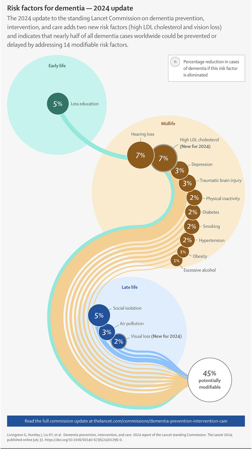
The Lancet Commission on dementia has just published its 2024 update on dementia prevention, intervention, and care. This report highlights multiple risk factors for dementia that are considered to be "modifiable" - issues that can be targeted to reduce risk. For the first time, the report included vision impairment on the list, which also includes less education, hearing loss, hypertension, smoking, obesity, depression, physical inactivity, diabetes, excessive alcohol consumption, traumatic brain injury, air pollution, and social isolation. Dr. Cecilia Lee was quoted in the New York Times article about the new Lancet report, and was also recognized in this UW Medicine news release: Vision loss, high cholesterol linked to higher dementia risk which highlights the Adult Changes in Thought study.
The Lancet report cites two studies by Cecilia Lee and co-authors as evidence for this new modifiable risk factor. This study on dementia outcomes after cataract extraction in the Adult Changes in Thought study cohort was a key piece of evidence connecting vision loss and increased risk of dementia development. Another study by Dr. Lee and her ACT colleagues that was cited in the Lancet report found that diabetic retinopathy, an ocular complication of diabetes, appears to be an important biomarker of dementia risk in people with type 2 diabetes.
Going forward, researchers will continue to investigate these connections between the eye and brain to better understand the mechanisms behind them. In the meantime, doctors and patients may consider interventions to counteract the hearing and vision loss that can often occur in older age. If nothing else, this can help prevent older people from becoming socially isolated - another modifiable risk factor for dementia.
This new paper, published in Nature Reviews Cardiology, reports findings and guidelines from a workshop conducted by the National Heart, Lung, and Blood Institute. The workshop aimed to identify the knowledge gaps and the opportunities for future research on using the eye as a window to systemic disease, including developing a roadmap for the use of retinal imaging biomarkers for cardiovascular disease risk prediction. The retina is well suited to this task because noninvasive retinal imaging technology is rapidly advancing and can provide a detailed assessment of the retinal vasculature. Structural and functional retinal vascular changes have been associated with cardiovascular disease as well as other. systemic diseases.
The workshop identified several roadblocks that must be addressed in order to facilitate the development of retinal imaging biomarkers of cardiovascular disease. One key challenge is the need to simplify and standardize the process for obtaining high quality images, especially by technicians and clinicians in non-ophthalmic settings. Another challenge is the lack of longitudinal studies in diverse participant groups - the large datasets needed for this sort of analysis can be complicated by ethical and patient privacy concerns. Many other research gaps were identified, including improving the measurement of structural and functional biomarkers and improved standardization of the imaging devices and protocols.
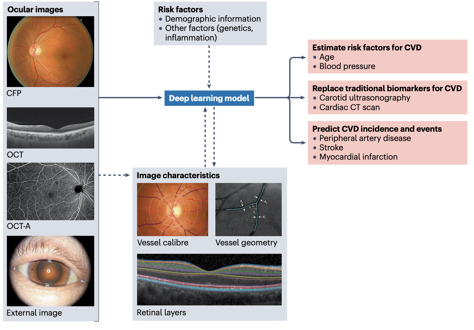
In addition, more research into the relationship between microvascular and macrovascular risk factors is critical, and there is increasing interest in combining retinal biomarker data with other data such as genetics and lifestyle-related risk factors using artificial intelligence approaches (see Figure).
Application of AI to retinal images. Ocular images, such as colour fundus photographs (CFPs), optical coherence tomography (OCT) and OCT angiography (OCT-A) scans, and external photographs of the eye are fed into deep learning models, either independently or together with information on other relevant risk factors to increase the predictive accuracy. Advanced image analysis techniques and deep learning models can extract meaningful features from these ocular images (such as vessel calibre, vessel geometry and retinal layers), which can also be fed into the prediction models. The deep learning models can then be used to estimate risk factors associated with cardiovascular disease (CVD), potentially replacing the use of traditional biomarkers of CVD derived from more expensive imaging of the cardiovascular system. Moreover, deep learning models can be used to predict the risk of specific CVD events. Adapted from ref. 132, Springer Nature Limited.
Future directions identified in the workshop include developing the strategy for clinical implementation of retinal vessel analysis in clinical practice, encouraging interdisciplinary collaboration, the development of networks and consortia to help facilitate such collaboration, and the development of a centralized data repository to enable the sharing of resources while building a strong evidence base. Going forward, the hope is that simple noninvasive eye imaging will be widely accepted as a key window into systemic health, providing clinicians with another tool to monitor their patients overall health.
The new study, published in the Journal of Alzheimer's Disease, describes the inaugural cohort of the Eye ACT study,, which follows older adults with the goal of identifying retinal biomarkers of Alzheimer's disease and related dementias. Data including visual function, retinal imaging, and retrospective eye health information is collected from participants, who will be followed over time for any changes in their visual data as well as for development of dementia.
Cecilia Lee is the principle investigator of EyeACT, and in the UW news article she describes a key component of the study: visiting the homes of certain participants who are unable to travel to a research clinic due to more complex health/mobility issues and collecting retinal imaging and clinical eye data from these patients using portable equipment. Previous work by the Adult Changes in Thought study (the parent study from which EyeACT recruits participants) has found that including data from older adults who unable to travel to a research clinic is essential to capturing the full picture of aging and dementia development.
EyeACT also collects retinal imaging and visual function assessments from patients who come into the clinic. The study will continue to follow these patients over time, but early examination of the initial data found that patients who were assessed at home had significantly worse visual function than those seen in clinic, highlighting the importance of including these patients to better understand the relationship between aging eyes and aging brains.
You can read more about the Eye ACT study in the recently published paper:
Lee CS, Ferguson AN, Gibbons LE, Walker R, Su YR, Krakauer C, Brush M, Kam J, Larson EB, Arterburn DE, Crane PK; Eye ACT Study Group:. Eye Adult Changes in Thought (Eye ACT) Study: Design and Report on the Inaugural Cohort. J Alzheimers Dis. 2024;100(1):309-320. doi: 10.3233/JAD-240203. PMID: 38875039.