Student becomes teacher: training faster deep learning lightweight networks for automated identification of optical coherence tomography B-scans of interest using a student-teacher framework

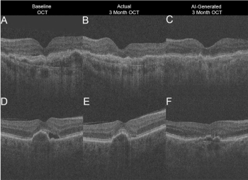
In this study, Dr. Julia Owen and her co-authors developed a novel method for automated detection of abnormal optical coherence tomography (OCT) B-scan ("B-scan of interest") images of the retina. Commercial OCT devices do not routinely provide automated diagnoses, so disease detection requires expert interpretation of the images by an ophthalmologist. Reliable automated detection of potentially abnormal retinal scans could save time, as the machine could flag abnormal scans that require further review by a clinician. But there have been challenges with developing such models, including the lack of large training datasets, standardized methods for image acquisition and processing, agreed-upon evaluation metrics, and limitations in computing power.

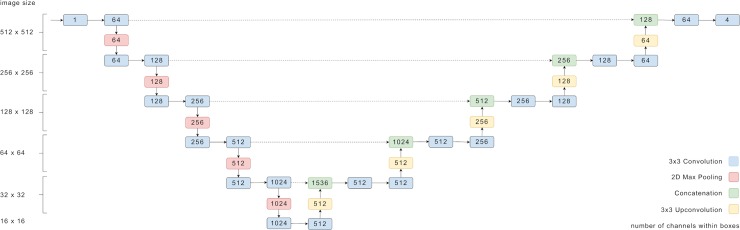
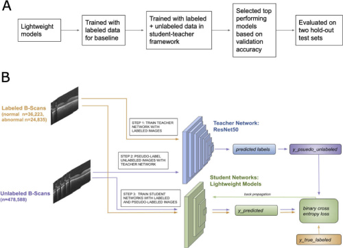
Dr. Owne developed an approach that overcomes some of these limitations, by using an efficient method for training lightweight machine learning models, or models with fewer parameters and operations, using unlabeled and routinely-available data. Lightweight models are useful in clinical settings because they can perform quickly and are more compatible with portable devices.

Continue reading "Student becomes teacher: training faster deep learning lightweight networks for automated identification of optical coherence tomography B-scans of interest using a student-teacher framework"

Methodological Challenges of Deep Learning in Optical Coherence Tomography for Retinal Diseases: A Review

In this review article for Translational Vision Science and Technology, Dr. Ryan Yanagihara and his co-authors discuss the progress and future directions of deep learning applications for diagnosing retinal disease from optical coherence tomography imaging.

Deep learning is a subfield of machine learning that involves convolutional neural networks. Within ophthalmology, deep learning has been applied to automated diagnosis, segmentation, big data analysis, and outcome predictions. Many recent studies have successfully used deep learning for OCT image analysis, in order to diagnose and segment features of diabetic retinopathy, age-related macular degeneration, and glaucoma.

Continue reading "Methodological Challenges of Deep Learning in Optical Coherence Tomography for Retinal Diseases: A Review"

Validation of automated artificial intelligence segmentation of optical coherence tomography images

Optical coherence tomography (OCT) is  one of the most rapidly-evolving imaging technologies used in ophthalmology.  OCT enables visualization of detailed anatomical structures in the eye.  Advances in artificial intelligence are simultaneously enabling technology that can learn to read these images and recognize features that are hallmarks for various diseases. Deep learning with artificial multi-layer neural networks has become very successful in these kinds of visual learning tasks.

Continue reading "Validation of automated artificial intelligence segmentation of optical coherence tomography images"

Generating retinal flow maps from structural optical coherence tomography with artificial intelligence

Advances in both imaging technology and artificial intelligence are enabling many new developments in the field of ophthalmology. Optical coherence tomography angiography (OCTA) is one such development. OCTA measures blood flow in retinal microvasculature and can visualize the superficial and deep capillary plexus of the retinal vasculature without the need for dye. But the technology is also expensive and has a limited field of view. In this study, we sought to develop a deep learning model that could address this issue by first learning to infer between structure and retinal vascular function from structural OCT images and then generating an OCTA-like image. This would allow for accurate objectively-generated annotations, eliminating the need for expert-defined labels, and potentially enable the acquisition of new information about retinal blood circulation from preexisting OCT databases.

Continue reading "Generating retinal flow maps from structural optical coherence tomography with artificial intelligence"

Use of Mechanical Turk as a MapReduce Framework for Macular OCT Segmentation

In this study published in the Journal of Ophthalmology, the authors experimented with distributing the work of performing a large complex image labeling task (identifying layers of the retina on multiple optical coherence tomography images) using a popular crowdsourcing marketplace. Optical coherence tomography is an important noninvasive diagnostic tool in the field of ophthalmology, especially for the management of age-related macular degeneration, a common cause of blindness in the developed world. However, the automated measurements provided by the optical coherence tomography software result in frequent errors for critical parameters such as macular thickness and volume. Human eyes have the ability to identify and complete areas on the image where there is poor signal to noise ratio and are better at recognizing complex pathology. The authors sought to evaluate the feasibility of using Mechanical Turk as a parallel platform to allow multiple people to simultaneously and rapidly perform manual segmentations (identification of retinal layer boundaries) of the images.

Continue reading "Use of Mechanical Turk as a MapReduce Framework for Macular OCT Segmentation"