

Optical coherence tomography (OCT) is one of the most rapidly-evolving imaging technologies used in ophthalmology. OCT enables visualization of detailed anatomical structures in the eye. Advances in artificial intelligence are simultaneously enabling technology that can learn to read these images and recognize features that are hallmarks for various diseases. Deep learning with artificial multi-layer neural networks has become very successful in these kinds of visual learning tasks.
These models can be trained not only to recognize and classify each pixel in an OCT image but to group them spatially and identify the groups as particular anatomical structures (semantic segmentation), and have been used to detect ophthalmologic conditions such as macular edema and to identify the layers of the retina. These are tasks that humans can also perform, of course, but require extensive training, can be time-consuming and tedious, and the results may vary between different human "graders." Machine learning models are capable of detecting pixel-level details that humans are not. The ability of these models to accurately identify particular eye structures at such a detailed level is a key task that will enable further advances in clinical medicine.
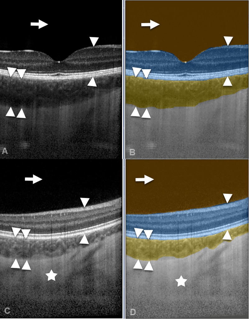
A spectral-domain OCT image (A) and a swept-source OCT image (C) were automatically segmented by the CNN (B, D) into the compartments vitreous (arrow), retina (arrow heads), choroid (double arrow heads), and sclera (asterisk).
In this study, a convolutional neural network (CNN) was developed to segment OCT images and the results were compared to different levels (non-medical, ophthalmologists, and research experts) of human graders. This study was intended to identify strengths and weaknesses of human graders compared to the CNN in identifying the vitreous, retina, choroid, and schlera compartments on the OCT images. The automated segmentation approach using a trained CNN achieved a more consistent result for all compartments at high scores comparable to human graders.
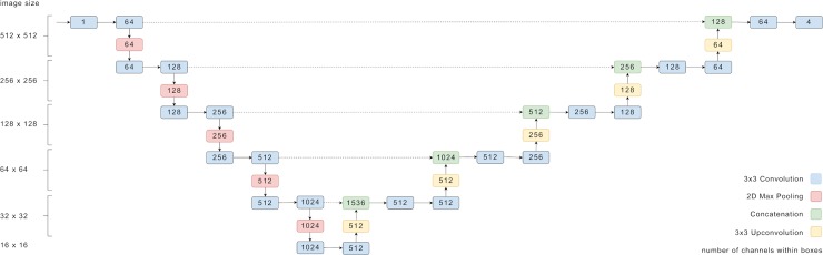
The numbers denote the dimensions of tensors passed between the layers.
This technology offers exciting potential for large-scale, high-quality OCT image compartmentalization for both research and patient monitoring purposes. This is an encouraging result, a step towards enabling automated detection of pathologies such as pigmented choroidal tumors. The developed method will be further enhanced and made available as a fully automated and cloud-based analysis method for pigmented choroidal tumors free of charge and open access to the medical community.
Maloca PM, Lee AY, de Carvalho ER, Okada M, Fasler K, Leung I, Hormann B, Kaiser P, Suter S, Hasler PW, Zarranz-Ventura J, Egan C, Heeren TFC, Balaskas K, Tufail A, Scholl HPN. Validation of automated artificial intelligence segmentation of optical coherence tomography images. PLoS One. 2019;14(8):e0220063. doi: 10.1371/ journal.pone.0220063. eCollection 2019. PubMed PMID: 31419240; PubMed Central PMCID: PMC6697318.