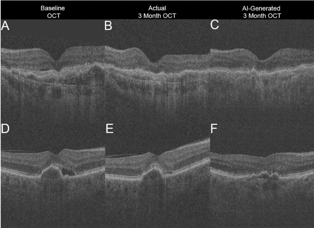
In this review article for Translational Vision Science and Technology, Dr. Ryan Yanagihara and his co-authors discuss the progress and future directions of deep learning applications for diagnosing retinal disease from optical coherence tomography imaging.
Deep learning is a subfield of machine learning that involves convolutional neural networks. Within ophthalmology, deep learning has been applied to automated diagnosis, segmentation, big data analysis, and outcome predictions. Many recent studies have successfully used deep learning for OCT image analysis, in order to diagnose and segment features of diabetic retinopathy, age-related macular degeneration, and glaucoma.
The authors note, however, that although deep learning has great potential as a diagnostic tool, numerous challenges have made it difficult to implement these algorithms into the clinical practice, including the lack of large datasets from multiple OCT devices, nonstandardized imaging and/or post-processing protocols between devices, limited graphics processing unit capabilities, and inconsistency in reporting metrics. The authors discuss these issues and possible ways to address them, as well as other general concerns that practitioners have about artificial intelligence and clinical practice.

They conclude that in order to best use this technology in the clinical practice of ophthalmology, a standardized framework for optical coherence tomography scans is necessary to increase the generalizability of deep learning analysis. Large, manually-annotated datasets using real patient data are also required to optimize the performance of these models to improve generalizability, and this method is superior to data augmentation and adversarialized image strategies. Although deep learning and OCT have individually revolutionized ophthalmology, optimizing the combined technologies will be integral to accelerating progress in the field.
Yanagihara RT, Lee CS, Ting DSW, Lee AY. Methodological Challenges of Deep Learning in Optical Coherence Tomography for Retinal Diseases: A Review. Trans. Vis. Sci. Tech. 2020;9(2):11. doi: https://doi.org/10.1167/tvst.9.2.11.

