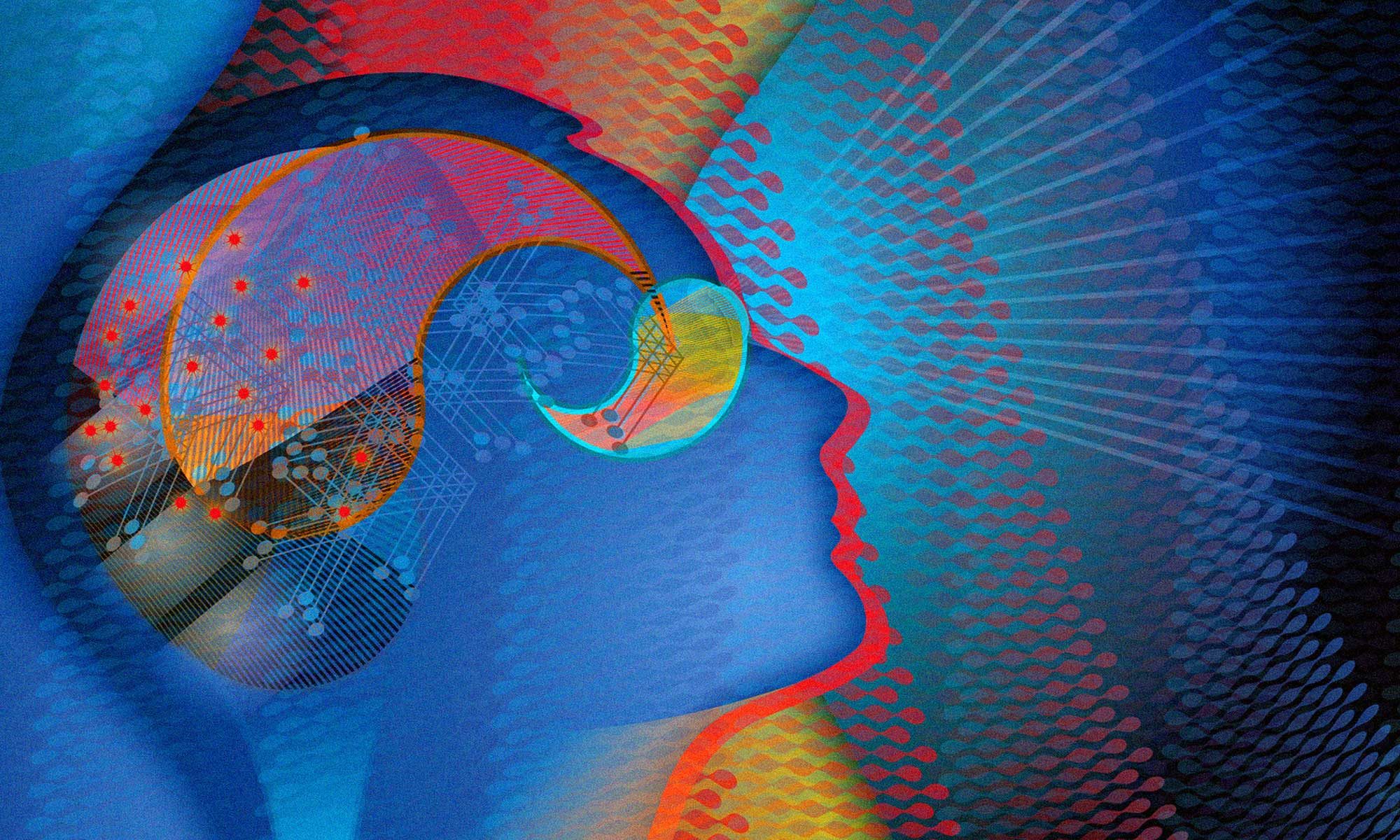
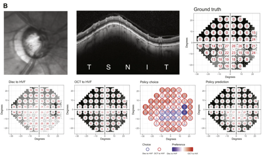
In a new paper published in the journal Ophthalmology, Lee Lab members Yuka Kihara and Aaron Lee report the development of a novel deep learning approach to address a key problem in glaucoma research and treatment - predicting visual function from structural changes detected on optical coherence tomography (OCT). Physicians rely on visual field testing to assess whether glaucoma patients will progress to more severe disease, but the test is notoriously challenging for patients and the results can be inconsistent. Advanced retinal imaging such as OCT can reveal key structural changes in the retina and optic nerve, but previous methods for linking structural changes to visual function outcomes have been challenging. This approach provides a fully automated method for improving the prediction of standard automated perimetry sensitivity directly from structural imaging data and also enables artificial intelligence–derived structure–function mapping.
Continue reading "Policy-Driven, Multimodal Deep Learning for Predicting Visual Fields from the Optic Disc and OCT Imaging"Association Between Cataract Extraction and Development of Dementia
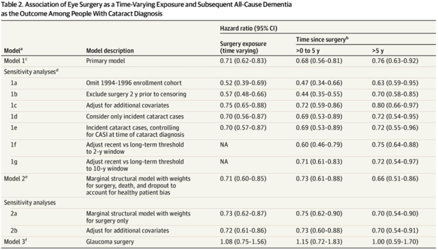
In a new paper published in JAMA Internal Medicine, Dr. Cecilia Lee and her colleagues report exciting findings related to the association between cataract surgery and dementia risk. Using data from Adult Changes in Thought, an ongoing longitudinal study following 5000+ older adults for the development of Alzheimer disease and other dementias, they compared outcomes of participants with cataract who had surgery to those who did not. Participants who underwent cataract surgery had nearly 30% lower risk of developing dementia from any cause compared with those who did not, even after controlling for many health-related confounders and potential sources of biases.
Continue reading "Association Between Cataract Extraction and Development of Dementia"Lee Lab Featured in Gates Notes
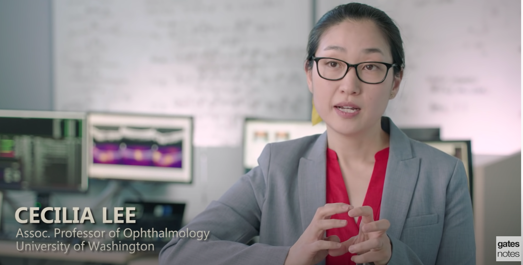
Several years ago, Bill Gates partnered with the Alzheimer’s Drug Discovery Foundation to develop a philanthropic fund called the Diagnostics Accelerator. Our own Dr. Cecilia Lee is one of the award recipients of this funding effort to discover and develop new diagnostic tests for Alzheimer's disease. Dr. Lee's lab is exploring retinal imaging techniques to identify early signs of Alzheimer’s, including by using artificial intelligence to analyze the imaging and potentially find irregularities that are invisible to the human eye. Bill Gates featured her research on developing retinal biomarkers for Alzheimer's disease in a recent Gates Notes blog post. Watch the video of Bill Gates describing this intiative and Dr. Lee discussing her research here.
Endophthalmitis Rate in Immediately Sequential versus Delayed Sequential Bilateral Cataract Surgery within the Intelligent Research in Sight (IRIS) Registry Data
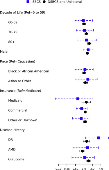
This large retrospective cohort study used data from more than 5 million people who underwent bilateral cataract surgery in the United States to investigate the rate of post-operative endophthalmitis. The authors compared the rates of endophthalmitis in the group of patients who had both eye surgeries on the same day (immediate sequential bilateral cataract surgery, or ISBCS) to the group of patients who either had cataract surgery in both eyes on separate days (delayed sequential bilateral cataract surgery) or who just had surgery in one eye. The authors found that after controlling for age, sex, race, insurance status, and comorbid eye diseases, the risk of postoperative endophthalmitis was not statistically significantly different between these two groups, .
Continue reading "Endophthalmitis Rate in Immediately Sequential versus Delayed Sequential Bilateral Cataract Surgery within the Intelligent Research in Sight (IRIS) Registry Data"Student becomes teacher: training faster deep learning lightweight networks for automated identification of optical coherence tomography B-scans of interest using a student-teacher framework
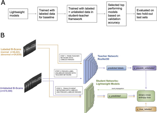
In this study, Dr. Julia Owen and her co-authors developed a novel method for automated detection of abnormal optical coherence tomography (OCT) B-scan ("B-scan of interest") images of the retina. Commercial OCT devices do not routinely provide automated diagnoses, so disease detection requires expert interpretation of the images by an ophthalmologist. Reliable automated detection of potentially abnormal retinal scans could save time, as the machine could flag abnormal scans that require further review by a clinician. But there have been challenges with developing such models, including the lack of large training datasets, standardized methods for image acquisition and processing, agreed-upon evaluation metrics, and limitations in computing power.
Dr. Owne developed an approach that overcomes some of these limitations, by using an efficient method for training lightweight machine learning models, or models with fewer parameters and operations, using unlabeled and routinely-available data. Lightweight models are useful in clinical settings because they can perform quickly and are more compatible with portable devices.
Continue reading "Student becomes teacher: training faster deep learning lightweight networks for automated identification of optical coherence tomography B-scans of interest using a student-teacher framework"