
The inflammatory maculopathies are a heterogeneous group of posterior uveitis syndromes that affect the macula. Most patients present with painless loss of vision with or without a viral prodrome, but differ in their clinical presentation, need for treatment, and visual prognosis. The rarity of these maculopathies and diverse spectrum of their presentation can make the diagnosis and management challenging. Fundus autofluorescence imaging provides additional information in a noninvasive manner. Inflammation may alter the quantity and the quality of the autofluorescence signal during the acute stage of macular disease. In this study, the authors aimed to develop a quantification method for fundus autofluorescence imaging, as well as describe the specific fundus autofluorescence features of these rare inflammatory maculopathies.
The authors reviewed the clinical findings, demographics, and fundus autofluorescence imaging characteristics of fifteen patients representing six different inflammatory maculopathies, and found differences between the various diseases. For example, two of the maculopathies, acute macular neuroretinopathy and unilateral acute idiopathic maculopathy, demonstrated the highest median autofluorescence values, suggesting global hyperautofluorescence during the active phase of the disease. The acute macular neuroretinopathy signal was characterized by overall loss of normal foveal hypoautofluorescence, however, while the unilateral acute idiopathic maculopathy showed a stippled and mixed pattern of hypo- and hyperautofluorescence. Although the classical visual description of these conditions may help to distinguish them clinically, fundus autofluorescence imaging may also be useful in distinguishing these disease syndromes in patients in whom findings may be more subtle or in patients who present later in their disease course.
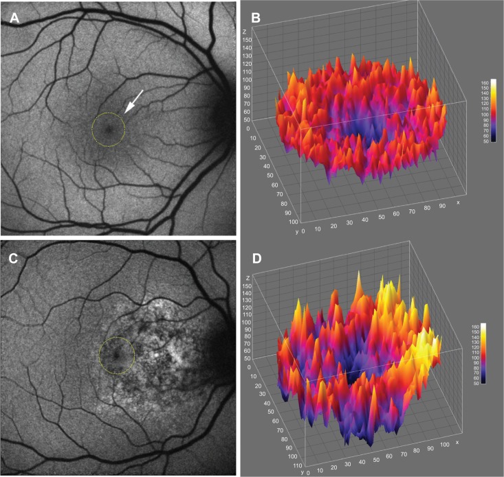
(A) FAF imaging of normal fundus. A circular area that corresponded to the size of the optic disc (white arrow) was placed on the center of the foveola (Indicated by the yellow circle). The median and standard deviation of foveal autofluorescence were measured in this area. (B) 3D heat map shows a 3D representation of low-to high-range FAF in the center of the foveola. (C) FAF in the acute stage of unilateral acute idiopathic maculopathy. The yellow circle corresponds to the center of the foveola, where the foveal autofluorescence was measured. (D) 3D heat map of the center of the foveola of a unilateral acute idiopathic maculopathy patient shows heterogeneous distribution of hyper- and hypoautofluorescence.
The authors were also able to specifically quantify autofluorescence for this rare group of inflammatory maculopathies, using medians and standard deviations to measusre foveal autofluorescence more objectively. The median autofluorescence value gives insight regarding the general hyper- or hypoautofluorescence of an image, while the standard deviation measures the diversity of the intensity distribution in a given region of interest. These measurements may be useful for providing quantifiable values that may be assessed over time as a disease process evolves, and may be a useful aid for distinguishing the various disease entities within this group.
Lee CS, Lee AY, Forooghian F, Bergstrom CS, Yan J, Yeh S. Fundus autofluorescence features in the inflammatory maculopathies. Clin Ophthalmol. 2014;8:2001-12. doi: 10.2147/OPTH.S68446. eCollection 2014. PubMed PMID: 25302012; PubMed Central PMCID: PMC4189704.