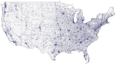
IgG4-associated orbital and ocular inflammation

IgG4-associated orbital and ocular inflammation is a relatively newly discovered disease which can manifest in the eye as sclerosing inflammation with infiltration of IgG4-positive plasma cells. This disease tends to affect tends to affect people in the middle-aged to elderly years, and the most commonly involved sites are the pancreas, the salivary and lacrimal glands, and the orbit. Some so-called idiopathic inflammation syndromes are being re-classified as IgG4-associated inflammation based on results from histopathologic evaluation. In this study, Dr. Lee and her coauthors report three cases of IgG4-associated ocular and orbital inflammation, each with a different presentation/manifestation: sclero-uveitis, pachymeningitis with associated bilateral optic neuropathy/perineuritis, and inflammatory pseudotumor of the orbit.

Continue reading "IgG4-associated orbital and ocular inflammation"

UK AMD EMR USERS GROUP REPORT V: benefits of initiating ranibizumab therapy for neovascular AMD in eyes with vision better than 6/12

Neovascular age-related macular degeneration is characterized by the growth of abnormal, choroidal blood vessels beneath the macula. When left untreated, these blood vessels can lead to severe loss of vision.  Intravitreal injection of anti-vascular endothelial growth factor-A drugs is an established therapy that helps to prevent the growth of these abnormal vessels. In the United Kingdom, this treatment is provided for patients with visual acuity in the range of 6/12-6/96, consistent with the pivotal trials that were conducted to approve the use of this therapy. As the use of intravitreal therapy for age-related macular degeneration has increased, studies have suggested that starting treatment earlier in the disease process, before patients have significant vision loss, can help to preserve vision longer. In this study, the authors used data from thousands of patients from multiple health centers in the United Kingdom to further evaluate this effect on a large scale.

Continue reading "UK AMD EMR USERS GROUP REPORT V: benefits of initiating ranibizumab therapy for neovascular AMD in eyes with vision better than 6/12"

Identification of torque teno virus in culture-negative endophthalmitis by representational deep DNA sequencing.

Although rare, infectious endophthalmitis is among the most serious post-surgical complications of ophthalmic surgery because it can result in serious vision loss. The standard technique for diagnosing endophthalmitis is microbial culture, but this technique fails to identify an organism in many cases. 16S polymerase chain reaction is more sensitive and specific than traditional culture techniques, but is limited to bacteria and cannot detect fungi (which require separate fungal rDNA ribosomal PCR), parasites, or viruses. Biome Representational in Silico Karyotyping (BRiSK) is a newer technique used to purify a defined fraction of all DNA present in a sample and sequence it to near saturation, allowing for the identification of most known bacteria, as well as phage, viruses, and previously unknown organisms. In this study, the authors applied this deep DNA sequencing technique to vitreous and aqueous biopsies from patients with endophthalmitis to test the hypothesis that uncultured organisms may be present in cases of culture-negative endophthalmitis.

Continue reading "Identification of torque teno virus in culture-negative endophthalmitis by representational deep DNA sequencing."

Fundus autofluorescence features in the inflammatory maculopathies

The inflammatory maculopathies are a heterogeneous group of posterior uveitis syndromes that affect the macula. Most patients present with painless loss of vision with or without a viral prodrome, but differ in their clinical presentation, need for treatment, and visual prognosis. The rarity of these maculopathies and diverse spectrum of their presentation can make the diagnosis and management challenging. Fundus autofluorescence imaging provides additional information in a noninvasive manner. Inflammation may alter the quantity and the quality of the autofluorescence signal during the acute stage of macular disease. In this study, the authors aimed to develop a quantification method for fundus autofluorescence imaging, as well as describe the specific fundus autofluorescence features of these rare inflammatory maculopathies.

Continue reading "Fundus autofluorescence features in the inflammatory maculopathies"