Data-Driven, Feature-Agnostic Deep Learning vs Retinal Nerve Fiber Layer Thickness for the Diagnosis of Glaucoma

In this commentary in the journal JAMA Ophthalmology, Christine A. Petersen, MD, Parmita Mehta, MS, and Aaron Y. Lee, MD, MSCI review a recent article published in the same journal, "Assessment of a segmentation-free deep learning algorithm for diagnosing glaucoma from optical coherence tomography scans."

The commentary addresses why a deep learning approach was more successful at detecting glaucoma using spectral domain optical coherence tomography scans than the more traditional strategy of performing automated segmentation to analyze retinal nerve fiber layer parameters.

Continue reading "Data-Driven, Feature-Agnostic Deep Learning vs Retinal Nerve Fiber Layer Thickness for the Diagnosis of Glaucoma"

Validation of automated artificial intelligence segmentation of optical coherence tomography images

Optical coherence tomography (OCT) is  one of the most rapidly-evolving imaging technologies used in ophthalmology.  OCT enables visualization of detailed anatomical structures in the eye.  Advances in artificial intelligence are simultaneously enabling technology that can learn to read these images and recognize features that are hallmarks for various diseases. Deep learning with artificial multi-layer neural networks has become very successful in these kinds of visual learning tasks.

Continue reading "Validation of automated artificial intelligence segmentation of optical coherence tomography images"

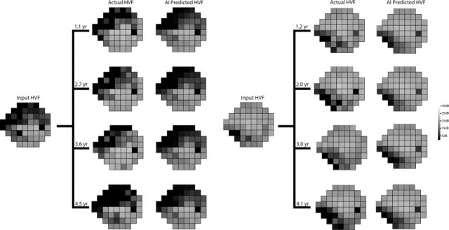
Forecasting future Humphrey Visual Fields using deep learning

Glaucoma is a leading cause of blindness worldwide, and the ability to quickly anticipate future disease progression may prevent unnecessary vision loss. Humphrey visual field testing is the standard method used to evaluate disease progression in glaucoma patients. This test uses linear regression of certain global eye measurements to monitor damage and predict risk for disease progression. It has some shortcomings in that does not take into account the spatial nature of visual field loss in glaucoma and relies on global indices rather than more focal measurements. Newer models have been developed that are better at detecting subtle changes and can identify disease progression earlier, but they require a multiple vision field tests to achieve accurate results. In this study, the authors aimed to develop a deep learning generative model to predict disease progression (to predict future visual fields with preserved spatial information) using minimal baseline visual field testing as input.

Continue reading "Forecasting future Humphrey Visual Fields using deep learning"

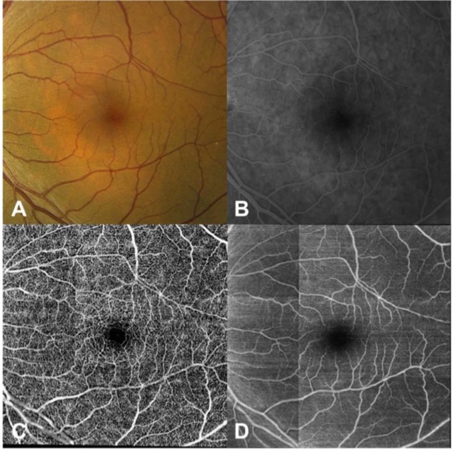
Generating retinal flow maps from structural optical coherence tomography with artificial intelligence

Advances in both imaging technology and artificial intelligence are enabling many new developments in the field of ophthalmology. Optical coherence tomography angiography (OCTA) is one such development. OCTA measures blood flow in retinal microvasculature and can visualize the superficial and deep capillary plexus of the retinal vasculature without the need for dye. But the technology is also expensive and has a limited field of view. In this study, we sought to develop a deep learning model that could address this issue by first learning to infer between structure and retinal vascular function from structural OCT images and then generating an OCTA-like image. This would allow for accurate objectively-generated annotations, eliminating the need for expert-defined labels, and potentially enable the acquisition of new information about retinal blood circulation from preexisting OCT databases.

Continue reading "Generating retinal flow maps from structural optical coherence tomography with artificial intelligence"

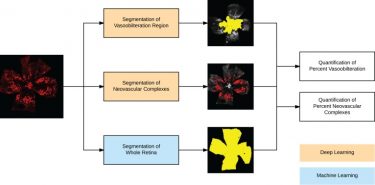
Fully automated, deep learning segmentation of oxygen-induced retinopathy images

In this paper, published in JCI Insight, the authors describe their deep learning model for quantifying vaso-obliteration and neovascularization on retinal images, which are key measurements in oxygen-induced retinopathy mouse model studies of ischemia-driven neovascularization. The mouse model of oxygen-induced retinopathy is one of the most commonly used in vivo models to study basic mechanisms in ocular angiogenesis and to test potential therapeutics. Measurements of vaso-obliteration and neovascularization are often used in proof-of-concept studies evaluating antiangiogenic drugs for diseases such as age-related macular degeneration and diabetic retinopathy.

Continue reading "Fully automated, deep learning segmentation of oxygen-induced retinopathy images"