
Optical coherence tomography (OCT) is the most commonly obtained imaging modality in ophthalmology and represents a dense and rich imaging dataset when combined with labels derived from the electronic medical record. In this study, the authors developed a novel deep learning approach to distinguish normal OCT images from images from patients with age-related macular degeneration.
OCT is used to monitor key pathologic findings in age-related macular degeneration, including drusen, retinal pigmented epithelium changes, and subretinal and intraretinal fluid deposits. Identification of these characteristics allows for precise management of neovascular age-related macular degeneration, and guides the decision of whether intravitreal therapy with anti-VEGF agents is needed. Computer aided diagnosis has the potential for allowing more efficient identification of pathological OCT images, as well as directing the attention of the clinician to regions of interest.
Deep learning is a subset of machine learning in which a many-layered neural network is trained to produce the desired results (distinguishing normal from diseased retinas in this case) purely from training data, without the need for labeled images. As a consequence, deep learning models require a large amount of training data. For this study, the authors extracted OCT images from a large imaging dataset and matched them with EHR data to determine whether or not patients had been diagnosed with age-relate macular degeneration. A total of 80,839 OCT images from patients with and without age-related macular degeneration were used for training the model, and 20,163 additional images were used for validation.
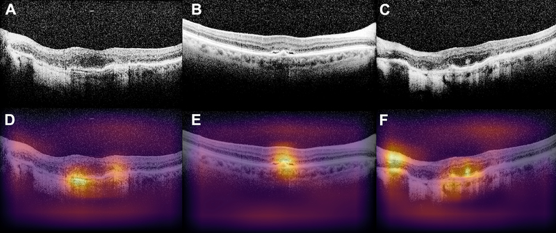
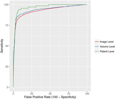
At the level of each individual image, the model achieved an accuracy of 87.63% with a sensitivity of 84.63% and a specificity of 91.54%. By averaging the probabilities from each image from the same patient, the model achieved an even greater accuracy (an area under the ROC curve of 97.46%). In addition, the authors used occlusion testing to determine which areas the model was identifying as regions of interest for age-related macular degeneration. The deep learning neural network successfully identified key areas which corresponded to known areas of pathology.
This deep learning algorithm provides a novel application to OCT classification in ophthalmology. This type of automated classification will likely be very beneficial for screening purposes, and the algorithm can also be used to identify concerning macular OCT images and efficiently display them to the clinician to aid in the diagnosis and treatment of macular pathology, much like the computer-aided diagnosis models found in radiology.
Lee CS, Baughman DM, Lee AY. Deep learning is effective for the classification of OCT images of normal versus Age-related Macular Degeneration. Ophthalmol Retina. 2017 Jul-Aug;1(4):322-327. doi: 10.1016/j.oret.2016.12.009. Epub 2017 Feb 13. PubMed PMID: 30693348; PubMed Central PMCID: PMC6347658.