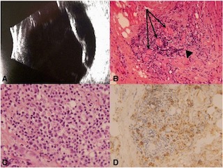
Identification of torque teno virus in culture-negative endophthalmitis by representational deep DNA sequencing.

Although rare, infectious endophthalmitis is among the most serious post-surgical complications of ophthalmic surgery because it can result in serious vision loss. The standard technique for diagnosing endophthalmitis is microbial culture, but this technique fails to identify an organism in many cases. 16S polymerase chain reaction is more sensitive and specific than traditional culture techniques, but is limited to bacteria and cannot detect fungi (which require separate fungal rDNA ribosomal PCR), parasites, or viruses. Biome Representational in Silico Karyotyping (BRiSK) is a newer technique used to purify a defined fraction of all DNA present in a sample and sequence it to near saturation, allowing for the identification of most known bacteria, as well as phage, viruses, and previously unknown organisms. In this study, the authors applied this deep DNA sequencing technique to vitreous and aqueous biopsies from patients with endophthalmitis to test the hypothesis that uncultured organisms may be present in cases of culture-negative endophthalmitis.

Continue reading "Identification of torque teno virus in culture-negative endophthalmitis by representational deep DNA sequencing."

Fundus autofluorescence features in the inflammatory maculopathies

The inflammatory maculopathies are a heterogeneous group of posterior uveitis syndromes that affect the macula. Most patients present with painless loss of vision with or without a viral prodrome, but differ in their clinical presentation, need for treatment, and visual prognosis. The rarity of these maculopathies and diverse spectrum of their presentation can make the diagnosis and management challenging. Fundus autofluorescence imaging provides additional information in a noninvasive manner. Inflammation may alter the quantity and the quality of the autofluorescence signal during the acute stage of macular disease. In this study, the authors aimed to develop a quantification method for fundus autofluorescence imaging, as well as describe the specific fundus autofluorescence features of these rare inflammatory maculopathies.

Continue reading "Fundus autofluorescence features in the inflammatory maculopathies"