Logistic Regression Classification of Primary Vitreoretinal Lymphoma Versus Uveitis by Interleukin 6 and Interleukin 10 Levels

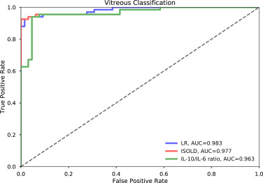
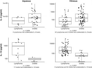
In this study published in the journal Ophthalmology, the authors evaluated the use of a logistic regression model for early diagnosis of primary vitreoretinal lymphoma. Primary vitreoretinal lymphoma is a rare disease with a generally poor prognosis. Early diagnosis of local ocular disease has been shown to prolong survival significantly, but because the disease is rare and the ocular symptoms are nonspecific, it is often misdiagnosed. In addition, although cytologic analysis of aqueous or vitreous samples is diagnostic, there are often problems with lymphoma cell detection in the sample. As a result, the diagnosis of PVRL is often delayed, taking on average 1 to 2 years from the onset of symptoms and typically requiring multiple biopsies.

Figure 4. Graph showing vitreous classification performance by interleukin 10 (IL-10)-to-interleukin 6 (IL-6) ratio, Interleukin Score for Intraocular Lymphoma Diagnosis (ISOLD), and logistic regression (LR). AUC = area under the receiver operating characteristic curve.
Continue reading "Logistic Regression Classification of Primary Vitreoretinal Lymphoma Versus Uveitis by Interleukin 6 and Interleukin 10 Levels"

Development and validation of a machine learning, smartphone-based tonometer

Access to eye care in more rural and remote areas can be challenging, because ophthalmologists need certain types of examination equipment that can be large and difficult to transport. Advances in smartphone technology are helping to address this problem - better cameras, software, and hardware attachments have the potential to allow ophthalmologists to bring a portable examining room to previously underserved areas.

Continue reading "Development and validation of a machine learning, smartphone-based tonometer"

Validation of automated artificial intelligence segmentation of optical coherence tomography images

Optical coherence tomography (OCT) is  one of the most rapidly-evolving imaging technologies used in ophthalmology.  OCT enables visualization of detailed anatomical structures in the eye.  Advances in artificial intelligence are simultaneously enabling technology that can learn to read these images and recognize features that are hallmarks for various diseases. Deep learning with artificial multi-layer neural networks has become very successful in these kinds of visual learning tasks.

Continue reading "Validation of automated artificial intelligence segmentation of optical coherence tomography images"

Medicare incentive payments to U.S. ophthalmologists for use of electronic health records: 2011-2016

The use of electronic health records offers a lot of advantages in terms of providing accurate, coordinated, up-to-date care for patients and may also help to reduce medical errors. The benefits depend on widespread implementation, however, and there have been challenges with getting physicians to use electronic records because they can require extra time, effort, and cost.

To encourage physicians to implement electronic health records into their practices, an incentive program was created by the Health Information Technology for Economic and Clinical Health (HITECH) Act of 2009, which provided direct payments yearly to physicians who adopted the use of electronic health records. To earn these incentive payments, providers were expected to demonstrate meaningful use of certified EHR technology according to metrics which were linked to certain goals for the program, such as improving efficiency and coordination of care, reducing disparities, etc. The program was designed to reward early adopters, as the payments decreased after the first two years.

Continue reading "Medicare incentive payments to U.S. ophthalmologists for use of electronic health records: 2011-2016"

Forecasting future Humphrey Visual Fields using deep learning

Glaucoma is a leading cause of blindness worldwide, and the ability to quickly anticipate future disease progression may prevent unnecessary vision loss. Humphrey visual field testing is the standard method used to evaluate disease progression in glaucoma patients. This test uses linear regression of certain global eye measurements to monitor damage and predict risk for disease progression. It has some shortcomings in that does not take into account the spatial nature of visual field loss in glaucoma and relies on global indices rather than more focal measurements. Newer models have been developed that are better at detecting subtle changes and can identify disease progression earlier, but they require a multiple vision field tests to achieve accurate results. In this study, the authors aimed to develop a deep learning generative model to predict disease progression (to predict future visual fields with preserved spatial information) using minimal baseline visual field testing as input.

Continue reading "Forecasting future Humphrey Visual Fields using deep learning"