
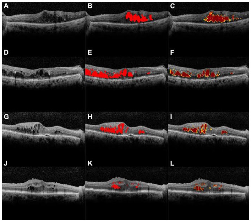
In this study, the authors developed a convolutional neural network that successfully detected intraretinal fluid on ocular coherence tomography images. Macular edema, characterized by loss of the blood retinal barrier in very small retinal blood vessels, leads to the accumulation of fluid in the retina that can interfere with vision. The degree of macula edema present is usually inferred by measuring central retinal thickness on ocular coherence tomography images, but this measurement does not fully capture the extent and severity of the intraretinal fluid present.
Manually identifying areas of intraretinal fluid on multiple images requires expertise, is extremely time consuming, and the results can vary between different experts. Prior studies have attempted automated segmentation (identification) of intraretinal fluid on ocular tomography images but were limited by the need for additional manual input. Some of these semi-automated methods have also had problems such as identifying areas incorrectly or not being about to distinguish between cystic and non-cystic fluid accumulations. To address these challenges, the authors used a novel deep learning approach to train a computer model to identify clinical features that are diagnostic of intraretinal fluid without the need for additional human input.
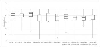
Using 1,289 ocular tomography images, the model segmented images with a 0.911 cross-validated Dice coefficient when compared with segmentations by human experts. The degree of agreement between model and the human experts was similar to the degree of agreement between the four independent clinicians. The deep learning approach used in this study showed promising results in accurate segmentation and automated quantification of intraretinal fluid volume on ocular coherence tomography imaging, and should be useful for future clinical and research applications.
Lee CS, Tyring AJ, Deruyter NP, Wu Y, Rokem A, Lee AY. Deep-learning based, automated segmentation of macular edema in optical coherence tomography. Biomed Opt Express. 2017 Jul 1;8(7):3440-3448. doi: 10.1364/BOE.8.003440. eCollection 2017 Jul 1. PubMed PMID: 28717579; PubMed Central PMCID: PMC5508840.