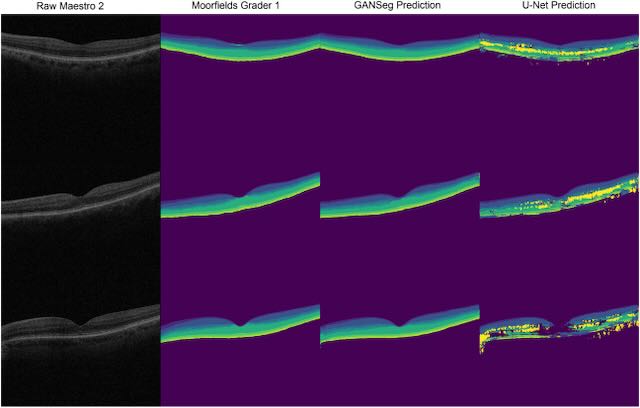
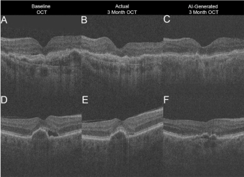
In this paper, recently published in the journal Ophthalmology, Yue Wu and his co-authors developed a unique method for training a model to identify the retinal layers on optical coherence tomography images, with the key feature that the model works successfully on optical coherence tomography (OCT) images from a different OCT device than the one that was used to obtain the training data. Although the field of deep learning image analysis is rapidly advancing, one major roadblock is the problem of domain shift, where models trained on a particular dataset may experience significant performance degradation when applied to slightly different datasets from different hospitals, imaging protocols, or device manufacturers.
Continue reading "Training Deep Learning Models to Work on Multiple Devices by Cross-Domain Learning with No Additional Annotations"Policy-Driven, Multimodal Deep Learning for Predicting Visual Fields from the Optic Disc and OCT Imaging
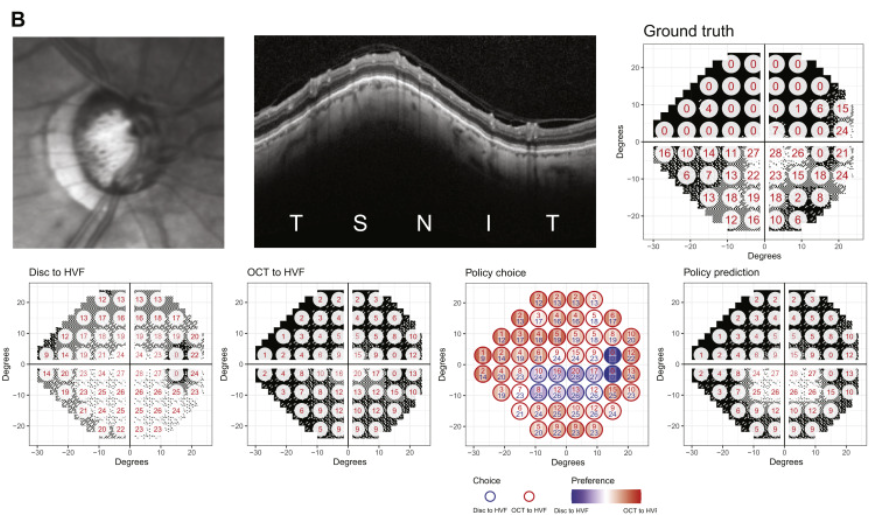
In a new paper published in the journal Ophthalmology, Lee Lab members Yuka Kihara and Aaron Lee report the development of a novel deep learning approach to address a key problem in glaucoma research and treatment - predicting visual function from structural changes detected on optical coherence tomography (OCT). Physicians rely on visual field testing to assess whether glaucoma patients will progress to more severe disease, but the test is notoriously challenging for patients and the results can be inconsistent. Advanced retinal imaging such as OCT can reveal key structural changes in the retina and optic nerve, but previous methods for linking structural changes to visual function outcomes have been challenging. This approach provides a fully automated method for improving the prediction of standard automated perimetry sensitivity directly from structural imaging data and also enables artificial intelligence–derived structure–function mapping.
Continue reading "Policy-Driven, Multimodal Deep Learning for Predicting Visual Fields from the Optic Disc and OCT Imaging"Application of deep learning to understand resilience to Alzheimer's disease pathology
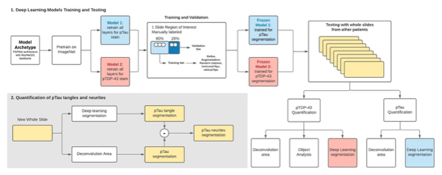
The term "resilient" is used to describe the unique set of people who develop the neuropathological features of Alzheimer's disease but do not show signs of the cognitive decline that is typically associated with the disease. In this study, published in the journal Brain Pathology, Dr. Cecilia Lee and Dr. Aaron Lee used a machine learning approach to identify subtle differences in brain tissue of "resilient" patients compared to patients with Alzheimer's disease. These patients had been participants in the Adult Changes in Thought study and donated their brains for research into the causes of Alzheimer's disease and related dementias.
Continue reading "Application of deep learning to understand resilience to Alzheimer's disease pathology"Using Deep Learning to Automate Goldmann Applanation Tonometry Readings
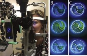
This study describes a novel method for automating Goldman Applanation Tonometry (GAT) measurements using a deep learning approach. GAT is the gold standard method for measuring intraocular pressure, which is an essential metric in the management of glaucoma.
To obtain accurate intraocular pressure measurements using traditional GAT, the user must use a dial to adjust the device to align two visible circular "mires", in order to determine the correct amount of force needed to create a fixed area of applanation on the surface of the eye, which is then used to calculate the intraocular pressure. This somewhat subjective alignment procedure can produce different results between users, and studies have found other unintended biases such as a preference for even-numbered readings on the dial. The goal of this study was to provide a method for obtaining more objective and reproducible GAT readings, by training a deep learning based algorithm to accurately recognize and measure the mires produced from a fixed application of force, and then use the measurements to calculate the intraocular pressure.
Continue reading "Using Deep Learning to Automate Goldmann Applanation Tonometry Readings"Methodological Challenges of Deep Learning in Optical Coherence Tomography for Retinal Diseases: A Review
In this review article for Translational Vision Science and Technology, Dr. Ryan Yanagihara and his co-authors discuss the progress and future directions of deep learning applications for diagnosing retinal disease from optical coherence tomography imaging.
Deep learning is a subfield of machine learning that involves convolutional neural networks. Within ophthalmology, deep learning has been applied to automated diagnosis, segmentation, big data analysis, and outcome predictions. Many recent studies have successfully used deep learning for OCT image analysis, in order to diagnose and segment features of diabetic retinopathy, age-related macular degeneration, and glaucoma.
Continue reading "Methodological Challenges of Deep Learning in Optical Coherence Tomography for Retinal Diseases: A Review"